1978 Confocal Laser Scanning Microscope

Confocal Laser Scanning Microscopy Clsm Of The Trunk Of Osedax Download Scientific Diagram

Neotorticaecum Kurochkin Nikolaeva 1978 From Paralichthys Download Scientific Diagram

Fluorescence Microscope

Laser Scanning Confocal Microscopy Images Of Salivary Gland Of 4 5 Download Scientific Diagram

Rp Photonics Encyclopedia Confocal Scanning Microscopes Operation Principle Pinhole Fluorescence Microscopy

Schematic Diagram Of A Confocal Laser Scanning Microscope Light Which Download Scientific Diagram
Unlike conventional optical microscopes these systems are mainly used for 3d surface analysis and characterization.
1978 confocal laser scanning microscope.

Light And Confocal Laser Scanning Microscopy Clsm Of Pyrodinium Download Scientific Diagram

Pdf Confocal Laser Microscopy Principles And Applications In Medicine Biology And The Food Sciences

Confocal Laser Scanning Microscopy And Scanning Electron Microscopy Of Tissue Ti Implant Interfaces Sciencedirect

Dual Channel Confocal Laser Scanning Microscope Photomicrographs Of Download Scientific Diagram

33 Laser Scanning Confocal Microscopy And Laser Microdissection Musculoskeletal Key

A Confocal Laser Scanning Microscopy Images For Inspecting The Download Scientific Diagram

Microscope History Timeline And The Application Of The Super Resolution Download Scientific Diagram

Confocal Laser Scanning Microscopy The Technique For Quantitative Fractographic Analysis Sciencedirect


Pteris Vittata A Young Gametophyte Without Mycorrhiza B Mature Download Scientific Diagram

Micro Ecology Of Peat Minimally Invasive Analysis Using Confocal Laser Scanning Microscopy Membrane Inlet Mass Spectrometry And Pcr Amplification Of Methanogen Specific Gene Sequences Sciencedirect

Figure 1 From Colocalization Of Cortical Microtubules And F Actin Indipodascus Magnusii Using Confocal Laser Scanning Microscopy Semantic Scholar

Laser Microscopy An Overview Sciencedirect Topics
Https Www Sciencedirect Com Science Article Pii 089106189190045e Pdf Md5 Fe534948843cfd1df4e5d9a0fc65a406 Pid 1 S2 0 089106189190045e Main Pdf

Milestones In The Development Of Two Photon Microscopy Download Table

Imaging The Cell And Molecular Dynamics Of Craniofacial Development Challenges And New Opportunities In Imaging Developmental Tissue Patterning Sciencedirect
Https Worldscientific Com Doi Pdf 10 1142 9789812790866 0057

Microscopic Photography Of Insects By Igor Siwanowicz Made With Confocal Laser Scanning Microscop Microscopic Photography Micro Photography Insect Photography
Https Encrypted Tbn0 Gstatic Com Images Q Tbn 3aand9gctpbujmtodhn0jsu29t6p4fqtrn7nyu8uhddir3vtckl5ocuqr Usqp Cau
Http Www Vacuumone Com Vacuumone Pdfs Newproducts Vacuumone Zetanoncontactopticalprofilers Pdf

Confocal Laser Scanning Microscope Clsm Micrographs Of Duck Egg Yolks Download Scientific Diagram

Pdf Scanning And Image Reconstruction Techniques In Confocal Laser Scanning Microscopy
Icfo On Twitter How Can Quantum Technology Improve Microscopy The Qmiceu Project Has Prepared An Infographic To Explain Why It Is A Completely New Approach By Implementing Quantum Imaging Technologies To Create

Testing Imaging Confocal Microscopy Laser Scanning Confocal Microscopy And Focus Variation Microscopy For Microscale Measurement Of Edge Cross Sections And Calculation Of Edge Curvature On Stone Tools Preliminary Results Sciencedirect

Confocal Microscopy An Overview Sciencedirect Topics

Hyphae Of Neurospora Crassa By Dr Eric Kalkman Plant Science Confocal Microscopy Laboratory Science

High Throughput Fret Analysis Of Protein Protein Interactions By Slide Based Imaging Laser Scanning Cytometry Szaloki 2013 Cytometry Part A Wiley Online Library

Pin On Spirals

Pdf Fractography Of Poly N Isopropylacrylamide Hydrogel Networks Crosslinked With Mechanofluorophores Using Confocal Laser Scanning Microscopy

Q Mic S New Infograph Qmic

Analyzing The Uf Resin Distribution In Particleboards By Confocal Laser Scanning Microscopy Sciencedirect
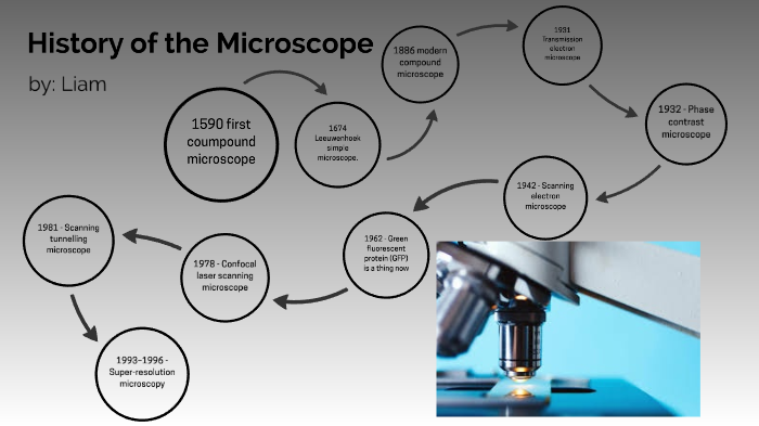
Microscopes By Liam Mail On Prezi Next

Imaging Confocal Microscopy Springerlink
Microscopy Presentation

Why Should Traceology Learn From Dental Microwear And Vice Versa Sciencedirect

Pdf Image Formation In Confocal Scanning Microscopes

Three Dimensional Imaging Of Monogenoidean Sclerites By Laser Scanning Confocal Fluorescence Microscopy
Https Www Jstor Org Stable 40058497

Pdf Study Of Skin Of An Egyptian Mummy Using A Scanning Electron Microscope
Https Ims Nus Edu Sg Events 2017 Data Files Sheppard Pdf

Pdf Spectral And Photobleaching Analysis Using Confocal Laser Scanning Microscopy A Comparison Of Modern And Archaeological Bone Fluorescence Tosha Dupras Academia Edu

Fluorescence Spectroscopy Of Ancient Sedimentary Organic Matter Via Confocal Laser Scanning Microscopy Clsm Sciencedirect
Https Asmedigitalcollection Asme Org Tribology Article Pdf 127 2 394 5698745 394 1 Pdf
Https Encrypted Tbn0 Gstatic Com Images Q Tbn 3aand9gcs8sytwgmkmgf6fmhtx9v0yupwtla Tc8ut 6vwtnyf131bovul Usqp Cau
Source : pinterest.com
